

Associated References
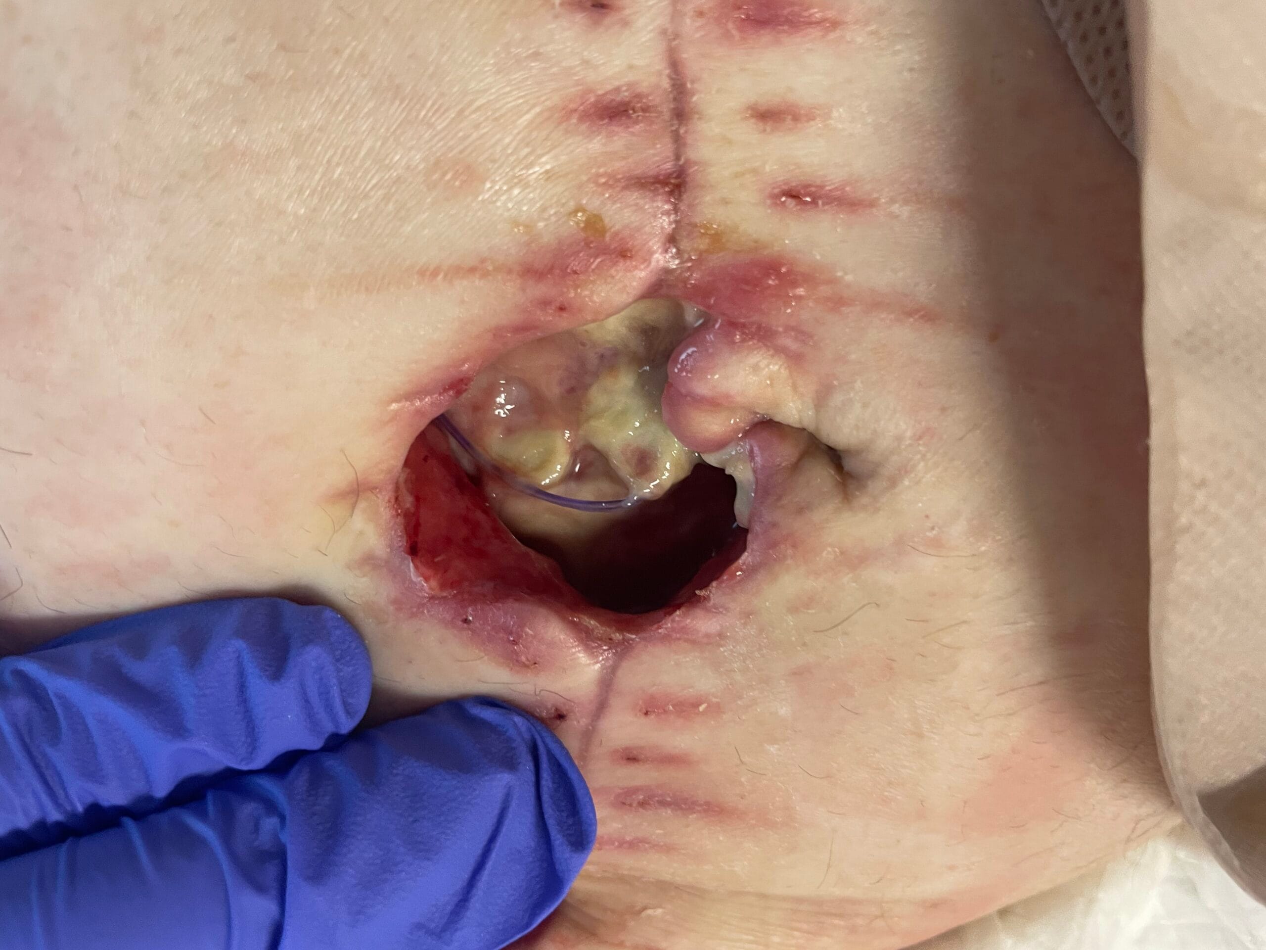
Photo Gallery
Main Article
Overview of Primary Closure Dehiscence
Primary closure dehiscence refers to the premature separation of the edges of a surgical wound that has been primarily closed. This condition can be classified into three categories: superficial, partial, and complete dehiscence. Superficial dehiscence involves only the skin layers, while partial dehiscence affects deeper layers, and complete dehiscence involves the full thickness of the abdominal wall or surgical site (Shahid et al., 2018; Gandhi et al., 2016). The common causes of primary closure dehiscence include infection, excessive tension on the wound, and systemic conditions such as obesity, diabetes, and malnutrition (Zoucas & Lydrup, 2014; Kajiwara et al., 2014). Risk factors associated with dehiscence are multifactorial, including surgical technique, patient comorbidities, and postoperative care practices (Raut et al., 2021; Shahid et al., 2022).
In surgical practice, the incidence of wound dehiscence varies significantly based on the type of surgery performed. For instance, emergency laparotomies have been reported to have a higher dehiscence rate compared to elective surgeries, with rates of 6.7% versus 1.5%, respectively (Gandhi et al., 2016). Furthermore, specific patient populations, such as those with complicated inflammatory diseases or neoplasms, exhibit even higher rates of dehiscence, emphasizing the need for tailored surgical approaches (Shahid et al., 2022; Sharma, 2018). Understanding these classifications and risk factors is crucial for healthcare professionals to implement effective preventive measures and management strategies.
Pathophysiology
The pathophysiology of wound dehiscence is complex and involves several physiological mechanisms that impair healing. Key factors include ischemia, infection, and compromised collagen synthesis. Ischemia can result from inadequate blood supply to the wound area, which is essential for delivering nutrients and oxygen necessary for healing (Althumairi et al., 2016; Surya, 2023). Infection is another critical factor, as it can lead to increased inflammation and tissue necrosis, further complicating the healing process (Chen et al., 2020; Lee et al., 2014).
Collagen synthesis is vital for wound healing, and any impairment in this process can lead to dehiscence. Conditions such as vitamin C deficiency, which is essential for collagen formation, can significantly affect wound healing (Meyer et al., 2015). Additionally, systemic conditions like diabetes can alter the inflammatory response and reduce the overall healing capacity of tissues (Elmarsafi et al., 2017; Liao et al., 2019). The interplay of these factors underscores the importance of a comprehensive understanding of wound healing mechanisms for effective management of primary closure dehiscence.
Acute Management
Acute management of primary closure dehiscence involves several evidence-based interventions aimed at assessing and addressing the wound’s condition. Initial wound assessment is critical, which includes evaluating the extent of dehiscence, presence of infection, and overall wound environment (O’Hanlan et al., 2016; Hyldig et al., 2016). Cleaning the wound is essential to remove any debris and necrotic tissue, which can harbor bacteria and impede healing (Tan et al., 2019). Debridement may be necessary in cases where necrotic tissue is present, as it promotes a healthier wound bed conducive to healing (Bellón et al., 2014; Shukla, 2024).
Techniques for re-closure vary based on the severity of dehiscence. Delayed primary closure is often recommended for cases where infection is present, allowing for adequate drainage and control of the infection before re-closure (Erdmann-Sager et al., 2018; Baka-Ostrowska et al., 2013). Secondary intention healing may be employed in cases where re-closure is not feasible, allowing the wound to heal from the bottom up (Ullah et al., 2021). Vacuum-assisted closure (VAC) therapy has also emerged as a beneficial technique, promoting granulation tissue formation and reducing the risk of infection (Nuveen et al., 2016; Seyrek & Akkuş, 2021). Each of these interventions should be guided by clinical evidence and tailored to the individual patient’s needs.
Long-Term Management
Long-term management strategies for preventing complications and promoting optimal healing following primary closure dehiscence are essential for improving patient outcomes. Scar management is a critical component, as hypertrophic scars can develop following dehiscence, impacting both function and aesthetics (Thrishuli & Kumar, 2018). Techniques such as silicone gel sheeting and pressure therapy have been shown to be effective in minimizing scar formation.
Nutrition optimization plays a vital role in wound healing, as adequate protein intake and micronutrient supplementation can enhance the healing process. Healthcare professionals should assess patients’ nutritional status and implement dietary interventions as needed. Infection control remains paramount, as postoperative infections can significantly hinder healing and lead to further complications. Regular monitoring and prompt intervention for any signs of infection are crucial components of long-term management strategies.
Precautions and Prevention
Preventing primary closure dehiscence requires a multifaceted approach that includes appropriate surgical techniques, thorough patient preparation, and diligent postoperative care. Surgical techniques should prioritize minimizing tension on the wound edges, utilizing techniques such as layered closure or continuous suturing, which have been shown to reduce dehiscence rates (Raut et al., 2021; Shahid et al., 2022). Patient preparation is equally important, involving preoperative optimization of comorbid conditions, such as controlling blood glucose levels in diabetic patients.
Postoperative care should include patient education on wound care and signs of infection, as well as regular follow-up appointments to monitor healing progress. The use of negative pressure wound therapy in high-risk patients has been shown to reduce the incidence of dehiscence and should be considered as a preventive measure (Hyldig et al., 2016; Thrishuli & Kumar, 2018). By implementing these strategies, healthcare professionals can significantly reduce the risk of primary closure dehiscence and improve overall surgical outcomes.
References:
- Althumairi, A., Canner, J., Gearhart, S., Safar, B., Fang, S., Wick, E., … & Efron, J. (2016). Risk factors for wound complications after abdominoperineal excision: analysis of the acs nsqip database. Colorectal Disease, 18(7). https://doi.org/10.1111/codi.13384
- Baka-Ostrowska, M., Kowalczyk, K., Felberg, K., & Wawer, Z. (2013). Pediatric urology complications after primary bladder exstrophy closure – role of pelvic osteotomy. Central European Journal of Urology, 65, 104-108. https://doi.org/10.5173/ceju.2013.01.art31
- Bellón, J., Pérez-López, P., Simón-Allué, R., Sotomayor, S., Pérez-Köhler, B., Peña, E., … & Calvo, B. (2014). New suture materials for midline laparotomy closure: an experimental study. BMC Surgery, 14(1). https://doi.org/10.1186/1471-2482-14-70
- Chen, H., Sheu, B., & Chang, W. (2020). Prolapsed epiploica of bowel after robotic hysterectomy: a case report. Annals of Medicine and Surgery, 60, 146-148. https://doi.org/10.1016/j.amsu.2020.10.055
- Elmarsafi, T., Garwood, C., Steinberg, J., Evans, K., Attinger, C., & Kim, P. (2017). Effect of semiquantitative culture results from complex host surgical wounds on dehiscence rates. Wound Repair and Regeneration, 25(2), 210-216. https://doi.org/10.1111/wrr.12509
- Erdmann-Sager, J., Wilkins, E., Pusic, A., Qi, J., Hamill, J., Kim, H., … & Chun, Y. (2018). Complications and patient-reported outcomes after abdominally based breast reconstruction: results of the mastectomy reconstruction outcomes consortium study. Plastic & Reconstructive Surgery, 141(2), 271-281. https://doi.org/10.1097/prs.0000000000004016
- Gandhi, J., Shinde, P., & Digarse, R. (2016). Evaluation of abdominal wall closure technique in emergency laparotomies at a tertiary care hospital. International Surgery Journal, 1796-1801. https://doi.org/10.18203/2349-2902.isj20162813
- Hyldig, N., Birke-Sørensen, H., Kruse, M., Vinter, C., Joergensen, J., Sørensen, J., … & Bille, C. (2016). Meta-analysis of negative-pressure wound therapy for closed surgical incisions. British Journal of Surgery, 103(5), 477-486. https://doi.org/10.1002/bjs.10084
- Kajiwara, K., Kimura, E., Nakano, M., Takano, H., & Okamoto, A. (2014). Clinical experience of j‐vac drain for skin closure in the laparotomy of obstetrics and gynecology. Journal of Obstetrics and Gynaecology Research, 40(4), 1089-1097. https://doi.org/10.1111/jog.12312
- Lee, M., Yang, H., & Kim, J. (2014). Reconstruction techniques for tissue defects formed after preauricular sinus excision. Archives of Plastic Surgery, 41(01), 45-49. https://doi.org/10.5999/aps.2014.41.1.45
- Liao, J., Chan, P., Cornwell, L., Tsai, P., Joo, J., Bakaeen, F., … & Chu, D. (2019). Feasibility of primary sternal plating for morbidly obese patients after cardiac surgery. Journal of Cardiothoracic Surgery, 14(1). https://doi.org/10.1186/s13019-019-0841-y
- Meyer, C., Diaz, A., Dalela, D., Hanske, J., Pucheril, D., Schmid, M., … & Trinh, Q. (2015). Wound dehiscence in a sample of 1 776 cystectomies: identification of predictors and implications for outcomes. Bju International, 117(6B). https://doi.org/10.1111/bju.13213
- Nuveen, E., Paek, P., & Nuveen, J. (2016). Patient satisfaction improves with delayed primary closure of dehiscence. The American Journal of Cosmetic Surgery, 33(1), 8-16. https://doi.org/10.1177/0748806816639022
- O’Hanlan, K., Emeney, P., Peters, A., Sten, M., McCutcheon, S., Struck, D., … & Hoang, J. (2016). Analysis of a standardized technique for laparoscopic cuff closure following 1924 total laparoscopic hysterectomies. Minimally Invasive Surgery, 2016, 1-9. https://doi.org/10.1155/2016/1372685
- Raut, V., Kumar, S., Raut, S., Bhate, K., Singh, M., Kakodkar, P., … & Waknis, P. (2021). Dehiscence rate in wound closed with cyanoacrylate and black braided silk after surgical removal of impacted third molar: a systematic review and meta‐ Oral Surgery, 15(1), 17-23. https://doi.org/10.1111/ors.12611
- Seyrek, Y. and Akkuş, M. (2021). What is the impact of a previously failed robicsek repair in the subsequent treatment of sternal dehiscence with thermoreactive nitinol clips?.. https://doi.org/10.22541/au.161780059.97703123/v1
- Shahid, M., Khan, F., Askri, Z., Asad, A., Alam, M., Ali, D., … & Afzal, M. (2022). One year of experience managing peritonitis secondary to gastrointestinal perforation at a tertiary care hospital: a retrospective analysis. Cureus. https://doi.org/10.7759/cureus.23966
- Shahid, M., Mahmoud, F., & Elmallah, S. (2018). Evaluation of a new technique for abdominal wall closure in midline laparotomies. International Surgery Journal, 5(8), 2701. https://doi.org/10.18203/2349-2902.isj20183188
- Sharma, D. (2018). Herring bone stitch: knitting to secure abdominal wall closure for emergency midline laparotomy. Gastroenterology medicine & research, 1(5). https://doi.org/10.31031/gmr.2018.01.000524
- Shukla, A. (2024). Herringbone stitch technique versus interrupted suturing technique for rectus closure in emergency laparotomy: a comparative study. Asian Journal of Medical Sciences, 15(4), 244-247. https://doi.org/10.3126/ajms.v15i4.62022
- Surya, R. (2023). Management of abdominal wound dehiscence following cesarean section in district area of indonesia: honey as an alternative dressing. Journal of South Asian Federation of Obstetrics and Gynaecology, 15(4), 472-474. https://doi.org/10.5005/jp-journals-10006-2280
- Tan, T., Rutges, J., Marion, T., Hunn, M., & Tee, J. (2019). Cyanoacrylate dermal closure in spine surgery: systematic review and pooled analysis. Global Spine Journal, 10(4), 493-498. https://doi.org/10.1177/2192568219861619
- Thrishuli, P. and Kumar, E. (2018). Abdominal wall closure in the presence of sepsis (acute abdomen): role of negative suction. International Surgery Journal, 5(2), 407. https://doi.org/10.18203/2349-2902.isj20180040
- Ullah, K., Uddin, S., Shoib, A., & Yaseen, M. (2021). Comparison of outcome of interrupted versus continuous closure technique of rectus sheath in emergency laparotomies patients in terms of wound dehiscence.. The Professional Medical Journal, 28(04), 455-458. https://doi.org/10.29309/tpmj/2021.28.04.6020
- Zoucas, E. and Lydrup, M. (2014). Hospital costs associated with surgical morbidity after elective colorectal procedures: a retrospective observational cohort study in 530 patients. Patient Safety in Surgery, 8(1), 2. https://doi.org/10.1186/1754-9493-8-2