

Associated References
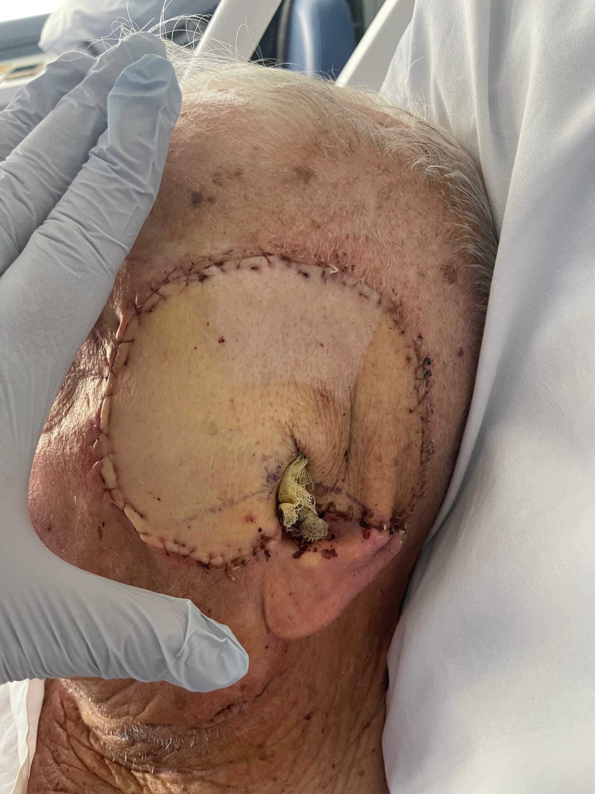
Photo Gallery


Main Article
Overview and History of Flap Techniques
Flap surgery, a cornerstone of reconstructive plastic surgery, has its roots in ancient practices, notably attributed to Sushruta, often referred to as the “Father of Plastic Surgery.” His work in the 6th century BC laid the groundwork for surgical techniques involving skin flaps for reconstructive purposes, particularly in nasal reconstruction (Bath et al., 2016). Over the centuries, advancements in surgical techniques and understanding of anatomy have significantly evolved flap surgery. The 20th century marked a pivotal era with the introduction of microvascular techniques, allowing for the transfer of free flaps, which are now a standard in reconstructive surgery (Jeong & Oh, 2016). Innovations such as perforator flaps, which utilize specific blood vessels to enhance flap viability while minimizing donor site morbidity, have further refined surgical approaches (Pignatti & Hallock, 2020).
The development of flap techniques has been driven by the need for effective reconstruction following trauma, oncological resections, and congenital deformities. The introduction of the free flap technique in the 1970s, particularly with the advent of microsurgery, revolutionized the field by enabling surgeons to transfer tissue from one part of the body to another with its blood supply intact (Jeong & Oh, 2016). This advancement allowed for more complex reconstructions, including those in the head and neck, where maintaining vascularity is crucial for flap survival (Jeong & Oh, 2016). The evolution of flap surgery continues with ongoing research into optimizing flap design and enhancing healing through biological agents such as growth factors (Fang et al., 2013).
Classification of Flaps
Flaps can be classified based on their vascular supply, composition, and method of transfer. The primary classifications include:
- Skin Flaps: These are the simplest form of flaps, consisting solely of skin and subcutaneous tissue. They are often used for covering defects in the skin, particularly in areas where minimal tension is required (Sanz & Simion, 2014).
- Muscle Flaps: These flaps consist primarily of muscle tissue and are used in situations where robust tissue coverage is needed, such as in the reconstruction of soft tissue defects after trauma or tumor excision (Kawamoto et al., 2018).
- Musculocutaneous Flaps: These flaps include both muscle and overlying skin, providing a versatile option for reconstructive needs. They are particularly useful in areas requiring both muscle bulk and skin coverage (Kawamoto et al., 2018).
- Fasciocutaneous Flaps: Comprising skin and fascia, these flaps are advantageous for their vascular supply and are often used in reconstructive surgeries where skin and underlying tissue need to be preserved (Sanz & Simion, 2014).
- Free Flaps: These flaps are detached from their original blood supply and reattached to a new site using microsurgical techniques. Free flaps are essential for complex reconstructions, especially in the head and neck region (Jeong & Oh, 2016).
- Axial and Random Pattern Flaps: Axial flaps are based on a specific artery, providing a reliable blood supply, while random pattern flaps rely on the surrounding vascular network, making them less predictable in terms of viability (Gavriilidis & Bota, 2019).
- Perforator Flaps: These flaps are based on perforating vessels that supply the skin and subcutaneous tissue. They can be categorized into direct and indirect perforator flaps, with direct perforators providing a more reliable blood supply. Various subtypes exist, including propeller flaps, which are rotated around a pivot point, allowing for versatile coverage options (Pignatti & Hallock, 2020; Ono, 2020).
Applications of Each Flap Type
The clinical applications of flap types vary significantly based on their characteristics and the specific needs of the reconstruction:
– Skin Flaps: Commonly used for wound coverage, skin flaps are essential in treating burns, traumatic injuries, and surgical defects. They provide a straightforward solution for superficial defects (Sanz & Simion, 2014).
– Muscle Flaps: These are particularly useful in reconstructing larger defects where muscle bulk is necessary, such as in the coverage of exposed bone or tendon (Kawamoto et al., 2018). They are often employed in oncological surgeries where extensive tissue removal has occurred (Ring et al., 2016).
– Musculocutaneous Flaps: These flaps are frequently used in head and neck reconstructions, where both skin and muscle are required to restore function and aesthetics (Jeong & Oh, 2016). Their versatility allows for the reconstruction of complex defects while maintaining a robust blood supply.
– Fasciocutaneous Flaps: These flaps are ideal for reconstructing defects in areas where skin quality is paramount, such as in the face or hands. They provide a balance between aesthetic outcomes and functional restoration (Sanz & Simion, 2014).
– Free Flaps: Free flaps are the gold standard for complex reconstructions, particularly in the head and neck, breast, and extremities. They allow for the transfer of tissue from distant sites while ensuring adequate blood supply, thus enhancing flap survival (Jeong & Oh, 2016).
– Axial and Random Pattern Flaps: Axial flaps are often used in reconstructive surgeries where a reliable blood supply is critical, such as in the reconstruction of the breast or after tumor excision (Gavriilidis & Bota, 2019). Random pattern flaps, while less predictable, can be useful in less complex reconstructions where the risk of ischemia is lower.
– Perforator Flaps: These flaps have gained popularity due to their ability to minimize donor site morbidity while providing adequate coverage. They are particularly useful in aesthetic surgeries and in areas where preserving underlying muscle is important (Pignatti & Hallock, 2020; Ono, 2020).
Surgical Techniques
The surgical techniques for flap procedures involve meticulous planning and execution to ensure optimal outcomes. Key considerations include:
- Door Site Selection: The choice of donor site is critical and should consider the flap’s vascular supply, the aesthetic implications, and the functional outcomes post-surgery. For instance, the radial forearm flap is commonly used for head and neck reconstructions due to its thin skin and reliable blood supply (Jeong & Oh, 2016).
- Harvest Techniques: Flap harvesting requires precision to preserve the vascular supply. Techniques vary based on flap type; for example, free flaps necessitate microsurgical techniques to anastomose the vessels at the recipient site, while local flaps may require simpler dissection (Jeong & Oh, 2016).
- Transfer Techniques: The transfer of flaps involves careful handling to maintain vascular integrity. In free flap procedures, the anastomosis of arteries and veins is performed under magnification to ensure patency and minimize complications (Jeong & Oh, 2016).
- Flap Design: The design of the flap should account for the defect’s size and shape, ensuring that the flap can adequately cover the area while maintaining tension-free closure at the donor site (Sanz & Simion, 2014).
- Intraoperative Monitoring: Continuous monitoring of flap perfusion during surgery is essential. Techniques such as Doppler ultrasound can be employed to assess blood flow to the flap (Jeong & Oh, 2016).
Postoperative Care and Monitoring
Postoperative care is crucial for the success of flap surgeries and involves several key components:
- Monitoring for Flap Viability: Clinicians must assess the flap for signs of viability, including color, temperature, and capillary refill. Any signs of ischemia, such as pallor or coolness, should prompt immediate intervention (Jeong & Oh, 2016).
- Prevention and Management of Complications: Complications such as ischemia, infection, and necrosis are significant concerns in flap surgeries. Prophylactic antibiotics may be administered, and careful wound care is essential to prevent infection (Hwang et al., 2018). Additionally, smoking cessation is advised prior to surgery to enhance healing (Hwang et al., 2018).
- Long-term Care Considerations: Postoperative care extends beyond immediate monitoring. Patients may require physical therapy to regain function, particularly in cases involving muscle flaps (Ring et al., 2016). Scar management techniques, including silicone gel sheets and massage, can help improve aesthetic outcomes (Sanz & Simion, 2014).
- Follow-up Protocols: Regular follow-up appointments are necessary to monitor the healing process and address any complications early. This includes assessing the donor site for healing and potential complications (Jeong & Oh, 2016).
- Patient Education: Educating patients about signs of complications and the importance of follow-up care is vital for successful outcomes. Patients should be informed about the healing process and what to expect post-surgery (Jeong & Oh, 2016).
Conclusion
Flap surgery represents a vital component of reconstructive plastic surgery, with a rich history and a diverse array of techniques tailored to meet the needs of various clinical scenarios. Understanding the classification, applications, surgical techniques, and postoperative care protocols is essential for healthcare professionals involved in managing skin and musculocutaneous flaps. Continued advancements in surgical techniques and postoperative care will further enhance the efficacy and safety of flap surgeries, ultimately improving patient outcomes.
References:
- Bath, K., Aggarwal, S., & Sharma, V. (2016). Sushruta: father of plastic surgery in benares. Journal of Medical Biography, 27(1), 2-3. https://doi.org/10.1177/0967772016643463
- Fang, T., Lineaweaver, W., Chen, M., Kisner, C., & Zhang, F. (2013). Effects of vascular endothelial growth factor on survival of surgical flaps: a review of experimental studies. Journal of Reconstructive Microsurgery, 30(01), 001-014. https://doi.org/10.1055/s-0033-1345429
- Gavriilidis, P. and Bota, E. (2019). Limberg flap versus karydakis flap for treating pilonidal sinus disease: a systematic review and meta-analysis. Canadian Journal of Surgery, 62(2), 131-138. https://doi.org/10.1503/cjs.003018
- Hwang, K., Son, J., & Ryu, W. (2018). Smoking and flap survival. Plastic Surgery, 26(4), 280-285. https://doi.org/10.1177/2292550317749509
- Jeong, W. and Oh, T. (2016). Oral and oropharyngeal reconstruction with a free flap. Archives of Craniofacial Surgery, 17(2), 45. https://doi.org/10.7181/acfs.2016.17.2.45
- Kawamoto, N., Okada, H., Hirohashi, K., Miyazaki, R., Yamamoto, M., Kume, M., … & Orihashi, K. (2018). Indocyanine green fluorescence/thermography evaluation of intercostal muscle flap vascularization. Thoracic Cancer, 9(12), 1631-1637. https://doi.org/10.1111/1759-7714.12871
- Ono, S. (2020). The history of propeller flaps. Seminars in Plastic Surgery, 34(03), 133-138. https://doi.org/10.1055/s-0040-1715157
- Pignatti, M. and Hallock, G. (2020). Introduction to “propeller flaps”. Seminars in Plastic Surgery, 34(03), 131-132. https://doi.org/10.1055/s-0040-1715156
- Ring, A., Kirchhoff, P., Goertz, O., Behr, B., Daigeler, A., Lehnhardt, M., … & Harati, K. (2016). Reconstruction of soft-tissue defects at the foot and ankle after oncological resection. Frontiers in Surgery, 3. https://doi.org/10.3389/fsurg.2016.00015
- Sanz, M. and Simion, M. (2014). Surgical techniques on periodontal plastic surgery and soft tissue regeneration: consensus report of group 3 of the 10th european workshop on periodontology. Journal of Clinical Periodontology, 41(s15). https://doi.org/10.1111/jcpe.12215